
Trying to evaluate for volume status or AAA in the long axis? Probe should be just subxiphoid with the indicator pointed to the patient’s head. For the IVC rock back and point the probe a bit towards the patient head. This should capture the right atrium in view. Identifying the heart and more specifically the right ventricle is an easy way to capture the IVC dumping into it. Here’s the distinguishing factors:
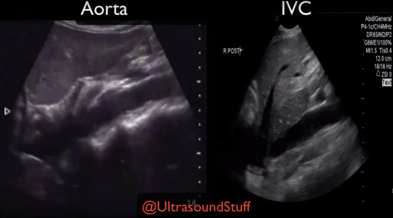
IVC: Thin walls, dumps into the right atrium and has the hepatic vein dumping into it just shy of the diaphragm, not as pulsatile as the aorta, is relatively to the right of the patient as compared to the aorta, and you may not see spine/vertebral bodies.
Aorta: Thick/hyperechoic walls, does not dump into the right atrium, can visualize the celiac and superior mesenteric arteries branching off proximally, the spine/vertebral bodies is typically readily apparent just posterior to the aorta, and it sits releativly left in the patient as compared to the IVC.

