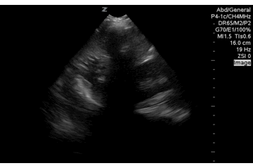

This image was mis-interpreted as a possible splenic injury during a FAST in the LUQ of the abdomen. If the user had pointed the ultrasound beam more posterior in the patient, to where dependent fluid should be they would notice the fluid filled stomach disappear out of view. Not free intraperitoneal fluid, but may be useful to note prior to intubation or if you are concerned for GI bleed.
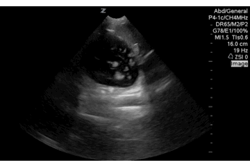
Another example fanning posterior and anterior. A better demonstration of rugae, contained fluid within the stomach and a negative LUQ FAST view.